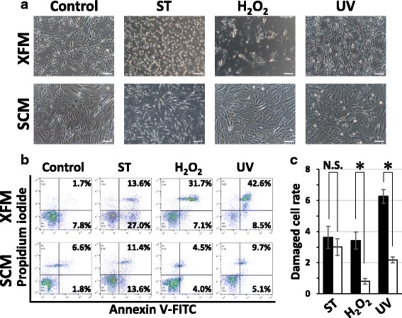
Fig. 3.

In-vitro assessment of cellular stress/damage of DPSCs induced by extrinsic cytotoxic stimuli under xenogeneic serum-free or FBS-containing culture conditions. a Morphological changes of DPSCs cultured in XFM and SCM before (control) and after treatment with staurosporine (ST), H2O2, or UV radiation. Scale bars, 100 μm. b Flow-cytometric analysis of cytotoxic stimulus-treated XFM and SCM cells using an Annexin V/PI system. c Quantification of the damaged cells cultured in XFM (black columns) and SCM (white columns). *P < 0.01. N.S. no significant difference, H2O2 hydrogen peroxide, UV ultraviolet, SCM xenogeneic serum-containing culture medium, XFM xenogeneic serum-free culture medium
