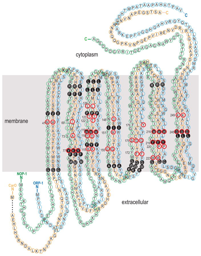
Figure 2.
Homology-based predicted secondary topology of the NOP-1 (green), ORP-1 (blue) and CarO (orange) protein in Fusarium graminearum. The seven transmembrane α-helices and helix boundaries are inferred by comparison to bacteriorhodopsin (Bieszke et al. 1999b; Grigorieff et al. 1996). Numbers indicate retinal-binding pocket residues that are conserved among the archaeal transport and sensory rhodopsins (Henderson et al. 1990; Hoff et al. 1997). A red circle around a numbered amino acid site indicates that the amino acid is shared with bacteriorhodopsin (i.e. the site is conserved). A solid black circle around a numbered amino acid site indicates that the amino acid is identical among the three fungal opsins.

