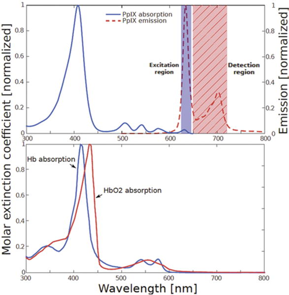
FIG. 1.

Upper: The portion of the PpIX absorption (excitation) spectrum corresponding to red illumination is indicated by the shaded rectangle labeled “Excitation region.” The portion of the PpIX emission spectrum detected during red-light illumination (the longer wavelength region of 650–720 nm) is indicated by the hatched rectangle labeled “Detection region.” Lower: The absorption spectra of deoxyhemoglobin (Hb) and oxyhemoglobin (HbO2) showing the strong absorption in the blue wavelength region that limits tissue penetration. Figure is available in color online only.
