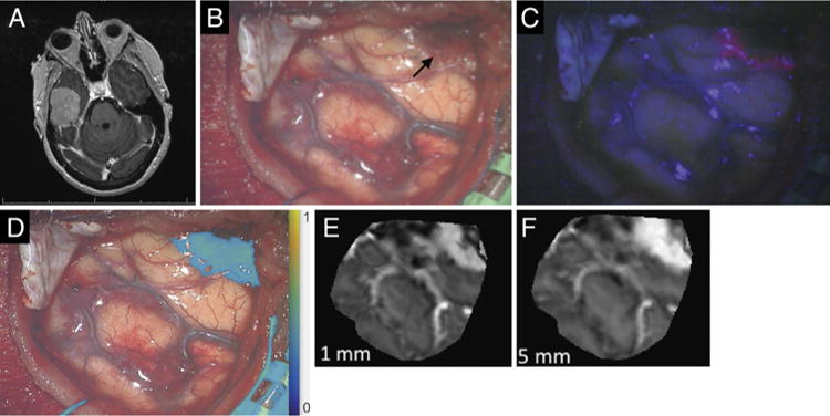
FIG. 5.

Case 11. A: Axial contrast-enhanced T1-weighted MR image of a well-demarcated tumor consistent with meningioma, arising from the floor of the right middle fossa. B: White-light image acquired upon initial exposure of the tumor (black arrow). C: Blue-light fluorescence image of the same surgical field. D: Overlay of fluorescence under red-light excitation. E: Reconstruction of MRI data corresponding to a weighted average of the first 1 mm below the surface of the surgical field. F: Reconstruction of weighted MRI data corresponding to the first 5 mm below the surface of the surgical field.
