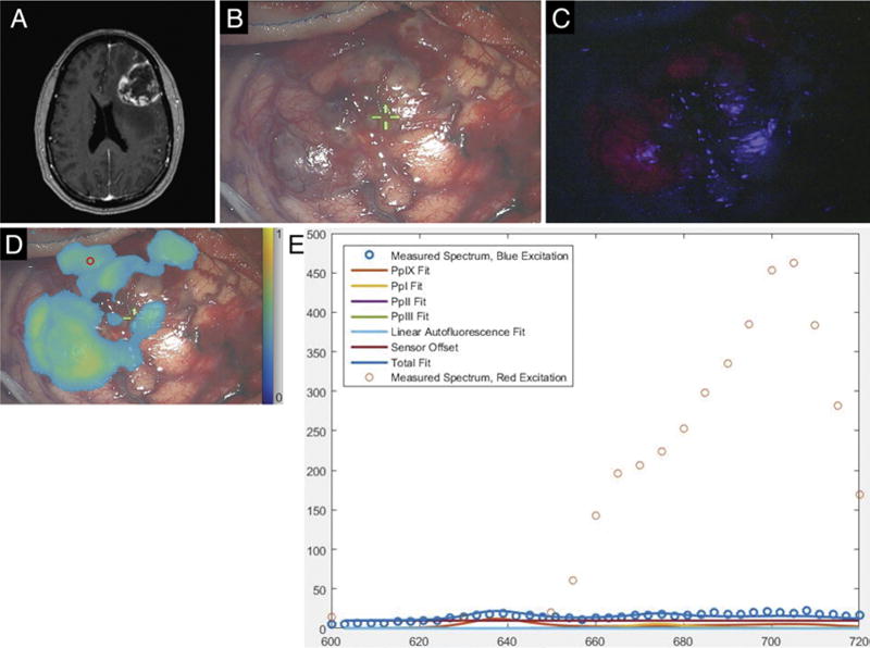
FIG. 6.

Case 24. A: Axial 3D MPRAGE image obtained after gadolinium administration, showing a heterogeneously contrast-enhancing, left frontal mass lesion. B: White-light image of tumor-involved cortical surface. C: Blue-light excitation fluorescence image of the same surgical field demonstrating fluorescence in areas of obvious tumor involvement. D: Map of PpIX fluorescence detected during red-light illumination superposed on a white-light image of the surgical field. The small red circle in the upper left portion of the image corresponds to the region of interest interrogated with the handheld optical probe. E: Optical spectra and model fits for PpIX, photoproducts, and autofluorescence at the site indicated by the small red circle in panel D. The typical blue light–elicited PpIX emission peak at 635 nm is absent (confirming the absence of conventional red fluorescence signal under blue-light excitation), while emission at longer wavelengths detected under red-light excitation confirms the presence of PpIX.
