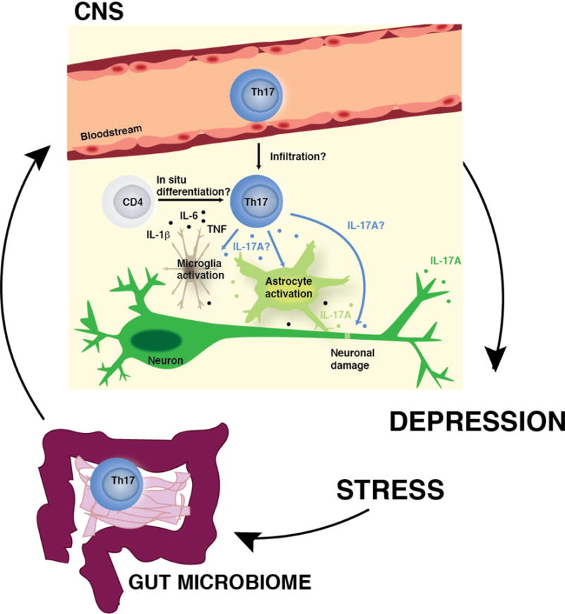
Figure.

Potential mechanisms of action of Th17 cells in the brain during depression. In response to stress, peripheral Th17 cells (possibly released from the lamina propria of the small intestine) may infiltrate the brain parenchyma, or surveying CD4+ T cells may differentiate into Th17 cells in situ after receiving signals from proinflammatory cytokines (e.g. IL-6, IL-1β, TNF) generated during the neuroinflammatory response to stress. Once in the brain parenchyma, Th17 cells may exert direct or indirect effects on the brain via production of IL-17A or other cytokines or factors produced by Th17 cells by either inducing activation of astrocytes and microglia, and/or by inducing neuronal damage, enhancing the neuroinflammatory response (including the production of IL-17A by CNS resident cells) and leading to increased susceptibility to depressive-like behaviors.
