Figure 1.

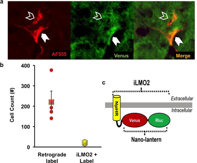
Detection of AAV-iLMO2 expression in the spinal cord. (a) Representative retrogradely labeled sciatic motoneurons with (solid arrowhead) and without (hollow arrowhead) Venus fluorescence, indicative of iLMO2 expression. (b) Quantification of detected iLMO2 in 10.7±3% of all retrogradely labeled sciatic motoneurons, (○) individual animal cell counts and (□) group mean ± SEM. (c) Schematic representation of iLMO2 (Tung et al. 2015).
