Fig. 1.

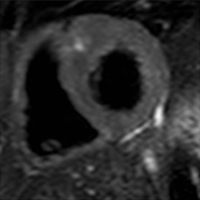
An imaging example of a HCM patient with HighT2. HighT2 was mostly demonstrated as a focal area in the hypertrophied anteroseptal wall at the insertion point of the right ventricle, as displayed here

An imaging example of a HCM patient with HighT2. HighT2 was mostly demonstrated as a focal area in the hypertrophied anteroseptal wall at the insertion point of the right ventricle, as displayed here