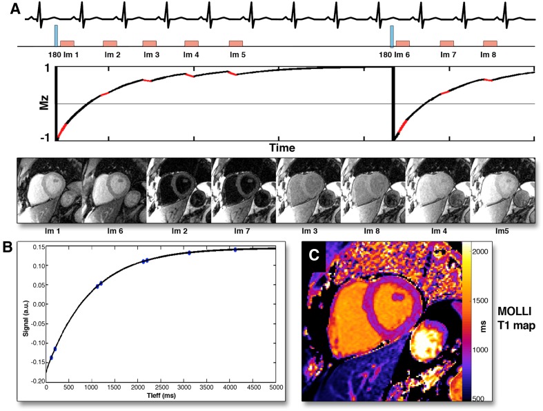
Fig. 1.
Modified Look-Locker (MOLLI) technique for myocardial T1 mapping. After radiofrequency inversion pulse, myocardial tissue longitudinal magnetization in a stable magnetic field returns to the equilibrium and a series of images are acquired in diastole over several heart beats (A). The images are sorted in order of increasing T1 times and the T1 recovery curve is obtained by plotting respective signal intensities against T1 time (B). The T1 map is obtained by applying this technique for all pixels in the image (C). Reproduced with permission from Taylor et al. [7]

