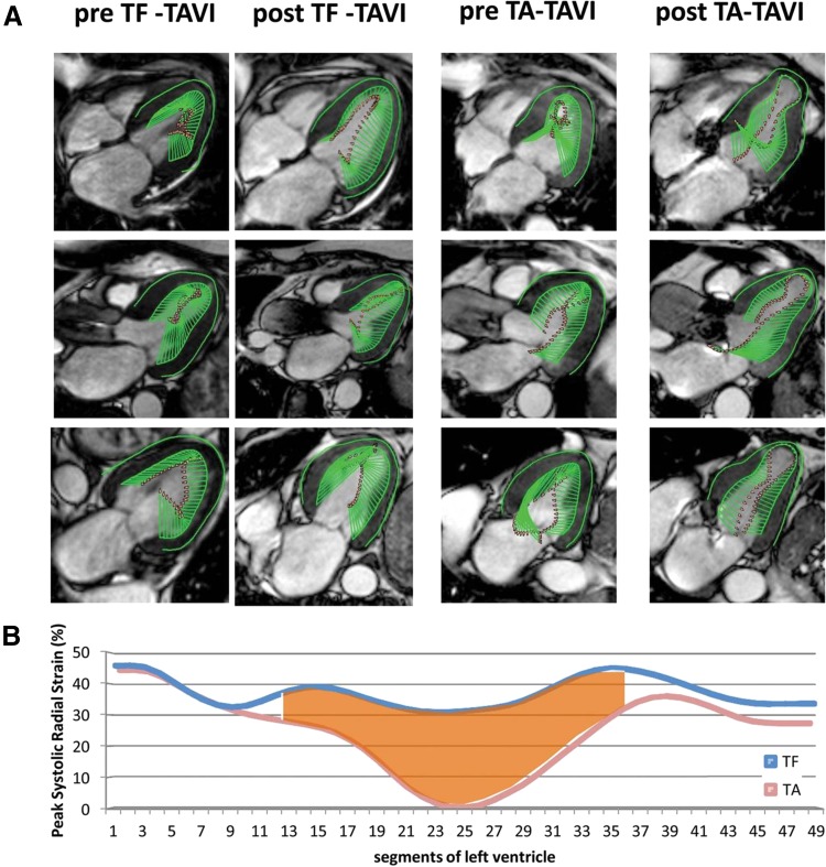
Fig. 7.
The impact of transcatheter aortic valve implantation on the left ventricular (LV) mechanics, assessed with feature tracking cardiovascular magnetic resonance (CMR). A Systolic CMR cine frames derived from four- (top row), three- (middle row), and two-chamber (bottom row) LV views of a patient before and after transfemoral (TF) access (left two columns) as well as from a patient before and after transapical (TA) access (right two columns). The green arrows represent velocity vectors illustrating systolic inward motion. The TA transcatheter aortic valve implantation (TAVI) patient shows reduced systolic deformation of the apical LV segments 3 months after the procedure. B Average peak systolic radial strain values of 49 analyzed segments obtained from all TF-TAVI patients (blue line) and all TA-TAVI patients (red line). The apical segments are displayed in the middle, while the basal segments are displayed on the left and on the right side of the graph. There is a reduction in peak radial strain of the apical segments after TA-TAVI. Adapted with permission from Meyer et al. [58]

