Figure 2.
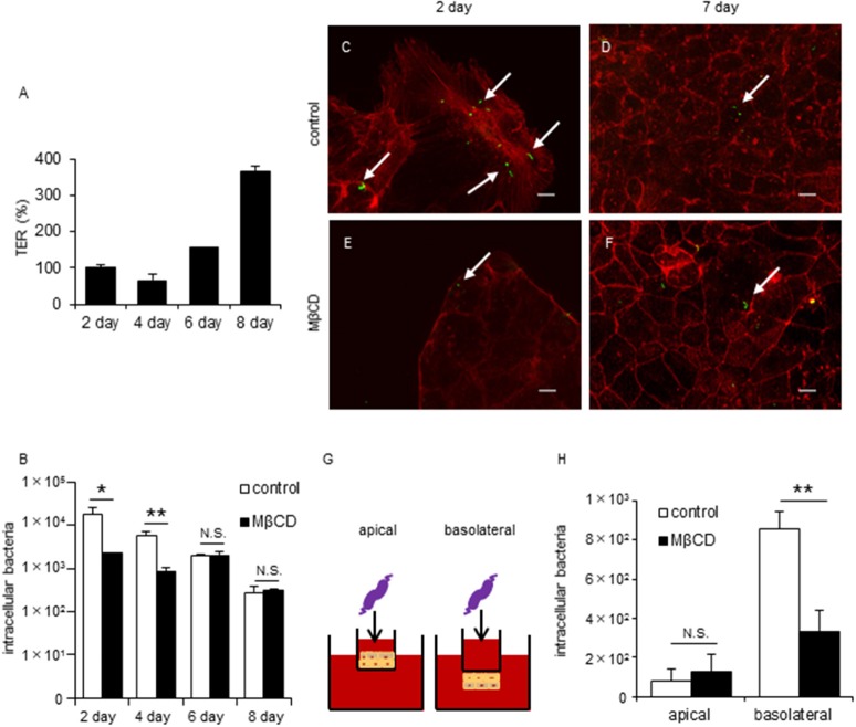
Disruption of lipid rafts reduces C. jejuni invasion of TJ unformed cells or infection of basolateral surfaces in polarized epithelial cells. Caco-2 cells were infected with C. jejuni for 6 h and the number of intracellular bacteria was measured by a gentamycin protection assay (B,H). (A) TER values were measured as a marker of TJ formation at 2, 4, 6, and 8 days post-seeding of Caco-2 cells cultured on transwells. TER values were calculated as the percentage of Day 2 post-seeding TER values. (B) Caco-2 cells cultured for 2, 4, 6, and 8 days following pre-treatment with 1-10 mM MβCD (black bar) or medium only (white bar) for 1 h. (C–F) Caco-2 cells cultured for 2 days (C,E) or 7 days (D,F) after pre-treatment with 1–10 mM MβCD for 1 h and subsequent infection with C. jejuni for 6 h. After infection, the cells were fixed, permeabilized and stained for F-actin (red). C. jejuni (green) was visualized using a CFDA SE cell tracer kit. Arrows indicate intracellular C. jejuni. Untreated control cells (C,D) and cells treated with MβCD alone (E,F) are also shown. Scale bar = 10 μm. (G,H) Caco-2 cells cultured in normal (apical) or inverted fashion (basolateral) on transwell inserts after pre-treatment with 10 mM MβCD for 1 h. Results are shown as the mean ± SD; n = 4. Significant difference from the control group are shown: N.S.; not significant; *P < 0.05; **P < 0.01.

