FIG. 5.

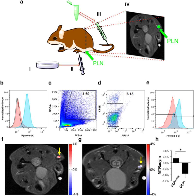
(a) Illustration of the experimental procedure. Ten million DCwt or DCDm-dNK activated with AddaVax Vaccine Adjuvant (InvivoGen, San Diego, CA, USA (1:1) were administered subcutaneously to the footpad of mice. Pyrrolo-dC was administered intravenously, 48 hours post-cell transplantation; and 2 hours postinjection, the PLNs were analyzed either by MRI or by FACS. (b) DCwt and DCDm-dNK were incubated in vitro with 4 mM pyrrolo-dC. Histogram displays the relative fluorescence of control DCwt+Pyrrolo-dC (red) and DCDm-dNK+Pyrrolo-dC (blue). Increased blue fluorescence corresponds to increased retention of pyrrolo-dC. No fluorescence was observed in the DCwt without pyrrolo-dC (empty histograms). (c-e) Ten million DCDm-dNK stained with CFSE (CFSE+ DCDm-dNK) were processed and injected into mice as described in (a), and PLNs were harvested and analyzed by FACS. The myeloid cell population was gated based on forward-scatter and side-scatter measure of granularity and size, respectively, (c) and the pyrrolo-dC uptake of the CFSE+ DCDm-dNK cells (d and e; blue) was compared to the CFSE− cells (APC-A channel is empty) (d and e; red). (f-h) Following the procedure described in (a), mice were imaged by MRI. CEST MRI map overlaid on anatomical MRI shows pyrrolo-dC accumulation in the PLN (arrow) of a mouse injected with DCDm-dNK (f; n = 5), but not with nontransduced DCwt (g; n = 5). (h) Average MTRasym values at 5.8 ppm plots of DCwt and DCDm-dNK (mean ± standared error of the mean; n = 5 mice per group), Student t test, unpaired two-tailed, P value = 0.046.CEST, chemical exchange saturation transfer; CFSE, carboxyfluorescein succinimidyl ester; dC, dendritic cells; Dm-dNK, drosophila melanogaster 2′-deoxynucleoside kinase; FACS, fluorescence-activated cell sorting; PLN, popliteal lymph nodes; pyrrolo-dC, pyrrolo-2′-deoxycytidine; SSC, side scatter; wt, wild type.
