FIGURE 2.

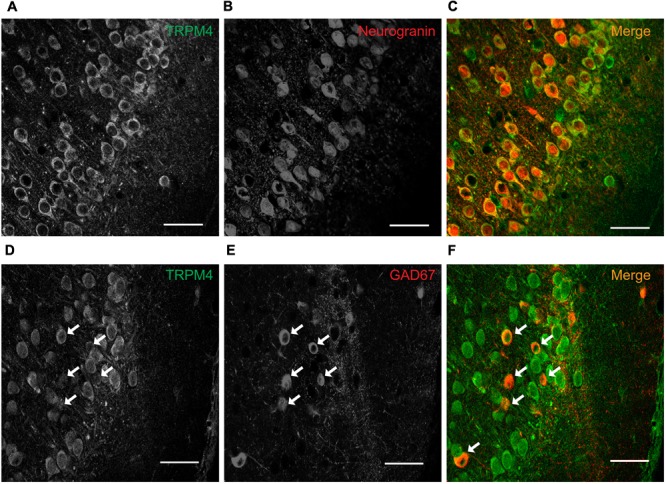
TRPM4 is expressed in pyramidal neurons and interneurons in layers 2/3 of the mPFC. Confocal images from double immunofluorescence labeling for (A) TRPM4 (green), (B) Neurogranin (red), and (C) the merged signals in a coronal mouse brain section at P35. Confocal images from double immunofluorescence labeling for (D) TRPM4 (green), (E) GAD67 (red), and (F) the merge of both signals in a coronal mouse brain section at P35. Arrows point to expression on interneurons. Scale bar = 40 μm.
