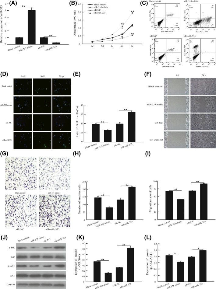
Figure 2.

miR‐335 inhibited cell growth, cell invasion and cell migration in vitro through the activation of the AKT/mTOR signaling pathway. A, A549 cells was transfected with exogenous miR‐335, miR‐335 antagomir or scrambled; the expression of miR‐335 was detected by quantitative RT‐PCR methods. B, Cell viability was assessed by MTT assay after transfection with different plasmids. C,D, A549 cells were transfected with miR‐335 siRNA, pre‐miR‐335 or negative controls for 24 h; then the cells were cultured with medium containing 10 μM BrdU for 1 h. Cells were fixed and stained for BrdU incorporation, immunofluorescence images of BrdU and DAPI were analyzed with Image J software and the ratio of BrdU‐positive cells was calculated. E, Cell apoptosis was detected by flow cytometric assay. F,G, Cell invasion was detected by transwell Matrigel assay, and number of invasion was measured with Image J software. H,I, Cell migration was detected by wound‐healing assay, and ratio of migration was measured with Photoshop CS5. J‐L, A549 cells were transfected with exogenous miR‐335, miR‐335 antagomir or scrambled for 48 h. Total proteins were extracted for immunoblotting of AKT, S6K, phosphorylation of AKT(S473) and S6K1(T389) and GAPDH. *P < .05 or **P < .01, vs pcDNA3.1 group. *P < .05 or **P < .01, vs ASO‐NC group
