
Figure 3.
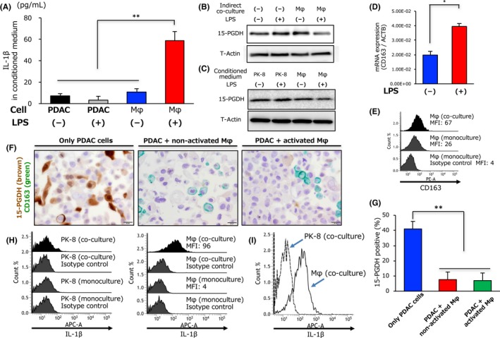
Activated tumor‐associated macrophages produce interleukin‐1β (IL‐1β) and reduce 15‐hydroxyprostaglandin dehydrogenase (15‐PGDH) expression in pancreatic ductal adenocarcinoma cells. A, Concentration of IL‐1β in conditioned medium from PK‐8 cells or macrophages (Mφ) treated with lipopolysaccharide (LPS) or distilled water (DW) was evaluated by ELISA. B, Expression of 15‐PGDH in PK‐8 cells determined by Western blot analysis 72 hours after co‐culture with or without macrophages treated with LPS or DW. C, Expression of 15‐PGDH in PK‐8 cells determined by Western blot analysis 72 hours after treatment with conditioned medium from PK‐8 or macrophages treated with LPS or DW. D, Quantitative RT‐PCR analysis of CD163 mRNA expression in macrophages 24 hours after LPS or DW treatment; data are presented as the LPS‐treated / control expression ratio. E, Expression levels of CD163 in monocultured macrophages (middle panel) or macrophages co‐cultured with PK‐8 cells (upper panel) were evaluated by flow cytometry. F, Representative double‐immunohistochemical staining of 15‐PGDH (brown) and CD163 (green) in only PK‐8 cells (left panel), PK‐8 cells directly co‐cultured with non‐activated macrophages (middle panel), or PK‐8 cells directly co‐cultured with activated macrophages (right panel). Scale bar = 20 μm. G, Column graph showing the percentage of 15‐PGDH‐positive cells in only PK‐8 cells, PK‐8 cells directly co‐cultured with non‐activated macrophages, or PK‐8 cells directly co‐cultured with activated macrophages. H,I, Expression levels of IL‐1β in monocultured PK‐8 cells or PK‐8 cells co‐cultured with macrophages and in macrophages co‐cultured with PK‐8 cells were evaluated by flow cytometry. **P < .01. APC‐A, allophycocyanin; MFI, mean fluorescence intensity; PE‐A, phycoerythrin