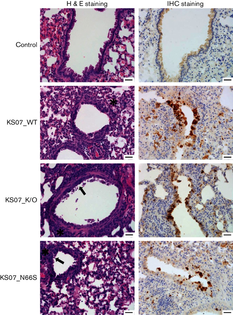
Fig. 4.

Haematoxylin and eosin and IHC staining of mouse lung sections at 7 days p.i. Haematoxylin-and-eosin-stained sections of mouse lungs infected with the indicated viruses at 7 days p.i. showed typical influenza pneumonia. Pictures are representative sections. In the control, no lesions are present, and there is no antigen deposition in the airway epithelium. In KS07_WT, there are areas of multifocal mild interstitial pneumonia (asterisk), and there is cuffing of bronchioles and vessels by lymphocytes and plasma cells. In KS07_K/O and KS07_N66S, there is mild epithelial hyperplasia (arrow), mild lymphocytic peribronchiolar cuffing (asterisk) and small amounts of fibrin and increased alveolar macrophages in alveolar lumina. IHC staining of lung sections was also conducted to detect influenza virus antigen using an anti-influenza A NP mAb (brown staining). All three treatment groups have positive antigen labelling for influenza NP in the nucleus of airway epithelial cells. Bars, 100 µm. H&E, Haematoxylin and eosin.
