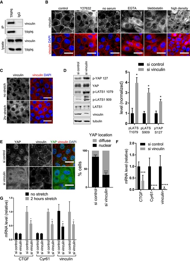
MCF10A cells were lysed, and anti‐TRIP6 or control (IgG) antibodies were used to isolate immune complexes. Immune complexes and lysates were probed by Western blotting for vinculin and TRIP6.
MCF10A cells were either not treated (control) or treated separately by growth to high density, serum starvation, Latrunculin B, Blebbistatin, EGTA, or Y27632 treatment and stained using anti‐vinculin antibodies by immunofluorescence. Scale bar = 20 μm.
MCF10A cells grown at high density on PDMS membranes and were stretched (or not) at 17% elongation for 2 h and fixed while under tension and stained for vinculin. Scale bar = 20 μm.
Vinculin was knocked down using two different siRNAs or control siRNA in MCF10A cells, and the cell lysates were probed by Western blotting for phospho‐LATS1 (T1079 and S909), phospho‐YAP (S127), LATS1, YAP, vinculin, and tubulin antibodies and the relative levels quantified (mean ± SD; n = 3; *P ≤ 0.05, t‐test).
Vinculin was knocked down as described in (D), and cells were stained for YAP and vinculin. Merged image shows YAP (green), vinculin (red), and DNA (blue). Quantification of at least 100 cells is shown (mean ± SD; n = 3; ***P ≤ 0.001, Fisher's test). Scale bar = 20 μm.
Vinculin was knocked down as described in (D), and the levels of vinculin and YAP target gene expression were analyzed using RT–qPCR (mean ± SD; n = 3; ***P ≤ 0.001, ****P ≤ 0.0001, t‐test).
Vinculin was knocked down in MCF10A cells as described in (D), grown at high density on PDMS membranes and cells were stretched (or not) at 17% elongation for 2 h and the levels of vinculin and YAP target gene expression were analyzed using RT–qPCR (mean ± SD; n = 3; *P ≤ 0.05, t‐test).

