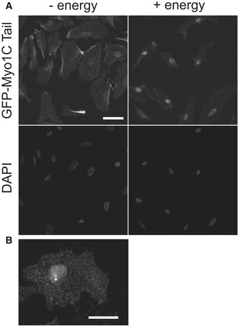
Figure 5. In vitro import assay shows that association of Myo1C with ER precedes its nuclear import.

- First, cells were incubated with import substrate (GFP‐Myo1C tail WT) in the absence of energy to saturate ER with the myosin (“− energy”). Then, unbound substrate was washed off and cells were supplemented with energy mix (creatine kinase, phosphocreatine, GTP, ATP), HeLa cytosolic extract, and incubated for 30 min at 30°C. Scale bar, 20 μm.
- A higher‐magnification image showing that recombinant GFP‐Myo1C tail WT associated with the membranes of the endoplasmic reticulum in digitonin‐permeabilized HeLa cells. Scale bar, 10 μm.
