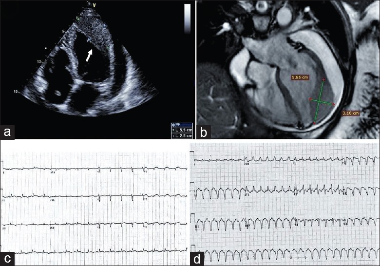
Figure 1.

(a) Transthoracic echocardiogram showed a new mass at the apex of the left ventricle and moderate-large pericardial effusion. The arrow points to the mass. (b) Cardiac magnetic resonance showed a homogeneous slightly hyperintense mass compared with the adjacent normal myocardium in the apical and lateral wall of the left ventricle and the pericardial effusion. (c) The 12-lead ECG on admission revealed sinus tachycardia with poor R wave progression in precordial leads and ST-segment elevation in leads V4 through V6. (d) The 12-lead ECG revealed ventricular tachycardia. ECG: Electrocardiogram.
