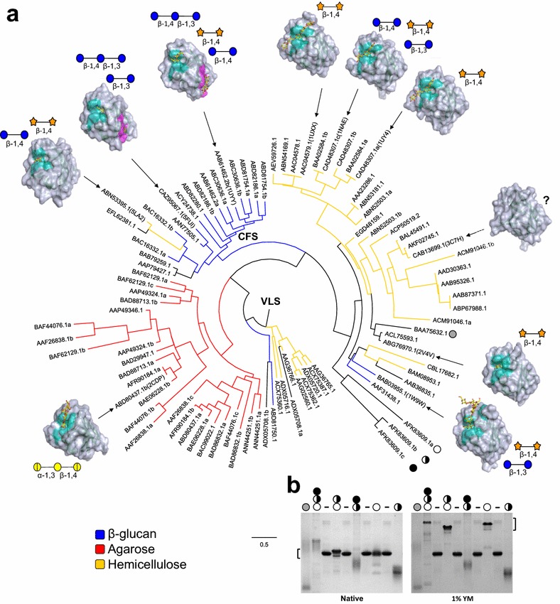
Fig. 3.

Distribution of CBM6 structures within a tree of CBM6s associated with characterized CAZymes. a Phylogenetic tree of characterized CBM6s (n = 90) were plotted with SACCHARIS. CBM6s with known three-dimensional structures where then mapped onto the tree and are indicated by their PDB ID. Rendered surface models with their bound ligands shown as yellow sticks are shown. For each structure the residues comprising the VLS are displayed in cyan and those of the CFS in magenta. Schematic representations of the sugar and stereochemical linkage recognized by each CBM are also displayed (blue circle = glucose, orange star = xylose, yellow circle = galactose, hatched yellow circle = 3,6-anhydro-l-galactose). Members of the tree are coloured based on the substrate that the appended catalytic module is active on. The black and white circles represent the CBM6s synthesized and tested for binding: BbCBM6 (grey circle), CcCBM6a (white circle), CcCBM6b (hatched circle), and CcCBM6c (black circle). b Affinity gel of various constructs of BbCBM6 and CcCBM6a–c. BSA controls are indicated with a dash. Equal amounts of CBM6s were run in acrylamide gels in absence (Native) and presence of 1% yeast mannan (YM)
