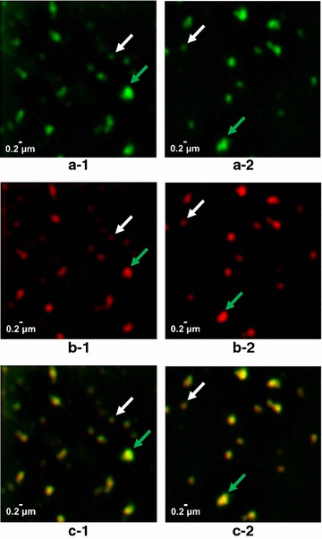
Fig. 9.

Chimeric VLP isolated from strain D#79 composed of dS and E2CSFV102-dS were analyzed under native conditions by N-SIM. Two series of images obtained from the same sample are presented showing fluorescence immunolabeling of dS in green (a-1, a-2), CSFV E2 antigen in red (b-1, b-2) and co-localization of the two labels in superimposed images in yellow (c-1, c-2). In each series of images two spots were consistently marked by arrows: signals of the size expected for individual VLP (white); largest signals in the respective frame (green)
