Fig. 1.

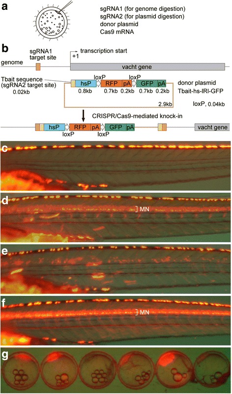
Strategy for the generation of knock-in medaka and generation of Tg[vacht-hs:lRl-GFP] strains. (a) For the generation of knock-in transgenic fish, sgRNA1 (for genome digestion), sgRNA2 (for plasmid digestion), donor plasmid with a bait sequence, and Cas9 mRNA are co-injected into one-cell-stage medaka embryos. (b) A schematic representation of the vacht locus (grey box) and the sgRNA target sites (orange box), and the reporter gene construct consisting of the Tbait (brown box), medaka hsp70 promoter (hsP, blue box), loxP, RFP-pA (red box), loxP, and GFP-pA (green box). After injection, the concurrent cleavage of the targeted genomic locus and the Tbait-hs-lRl-GFP reporter plasmid results in the integration of the reporter by non-homologous end joining (NHEJ). The scheme shows the forward integration of the reporter. (c) Lateral view (red fluorescence) of a control larva at 9dpf. Red-yellow signals in the dorsal region of the body are from the auto-fluorescence of pigment cells. (d) An example of an injected larva. RFP expression was present broadly in the motoneurons (MN). Animals with this kind of RFP expression were judged as having “good expression”, and raised to adulthood. (e) Another example of an injected larva. In this animal, RFP expression was present in the motoneurons, but the number of RFP-expressing cells is much smaller than that in (d). Animals with this kind of RFP expression were judged as not having “good expression”, and were not raised. (f) Lateral view of a Tg[vacht-hs:lRl-GFP] larva. RFP expression was present broadly in the trunk motoneurons. All of the trunk motoneurons are likely to express RFP in this animal. (g) Maternal expression of RFP in early embryos in the Tg[vacht-hs:lRl-GFP] line. The expression levels of RFP are variable among embryos. The embryos were obtained from a single mother
