
Figure 1.
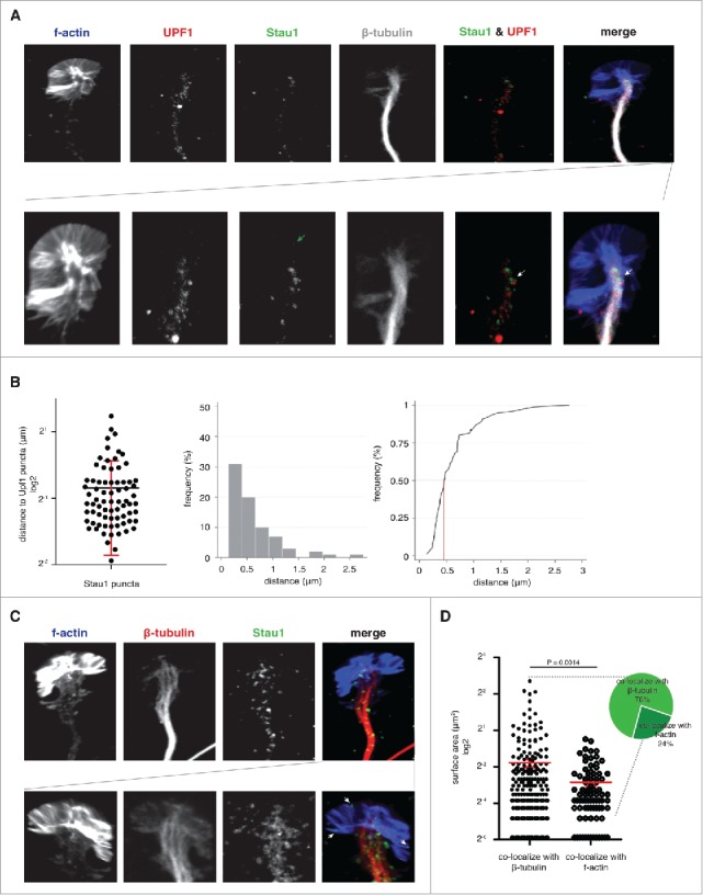
Staufen1 is detected near UPF1 in the mouse DRG axons and growth cones. A. Staufen1 is localized near UPF1 in the DRG growth cone. E14 DRG neurons were cultured for 4 DIV prior to staining to detect F-actin, Staufen1, UPF-1, and β-tubulin. The confocal images illustrate Staufen1 and UPF1's location within a DRG growth cone. Below is a magnified image of the growth cone to help visualize Staufen1 and UPF1 puncta. White arrows point to Staufen1 near UPF1 with a partial overlap. B. Half of Staufen1 puncta are located < 0.5 μm away from UPF1. The distance between Staufen1 and UPF1 was measured with ImageJ (n = 75) and plotted (left image) and represented as a histogram (middle image). A frequency distribution (right image) analysis shows that the majority of Staufen1 puncta are within 0.5 µm of a UPF1 puncta. The proximity of Staufen1 and UPF1 may be consistent with their interaction in SMD. C. Staufen1 particles show different sizes in axons compared to growth cones. Staufen1 and β-tubulin were detected by immunofluorescence and F-actin was detected using a CytoPainter F-actin staining kit (Abcam). Staufen1 puncta can be seen in axons in addition to growth cones. In many cases, Staufen1 puncta in growth cones were smaller than puncta in axons. White arrows point to examples of Staufen1 located at the distal ends of the growth cones. D. Average size of Staufen1-containing granules. Each dot represents a granule. Each granule was measured using ImageJ (n = 334 granules). The plot shows that the average size of the granules differs depending on their association with different cytoskeleton. Specifically, the granules at the distal ends are smaller than the granules close to β-tubulin.