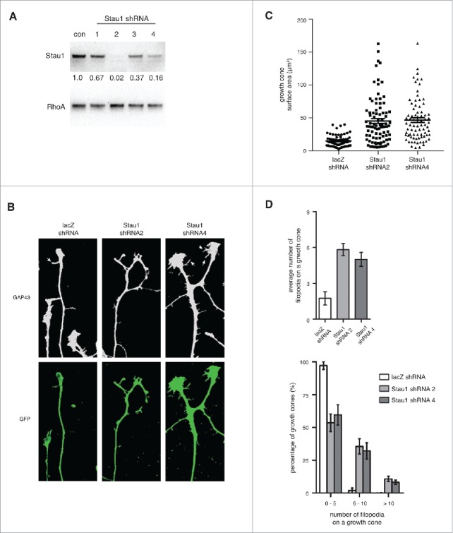
Figure 2.

Depleting Staufen1 causes abnormal DRG growth cone morphologies. A. Staufen1 was depleted in neurons using a lentivirus-mediated shRNA expression. Lysates were prepared from the control and Staufen1-depleted neurons and Staufen1 was detected by immunoblotting. About 98% and 80% of endogenous Staufen1 was depleted by shRNA2 and shRNA4, respectively. B. Growth cones show changes in morphology following Staufen1 depletion. DRG neurons were cultured for two days. Then, shRNA against lacZ (control) and Staufen1 was delivered to the neurons by lentivirus infection. After removing the lentivirus, the neurons were cultured for additional four days. The neurons were fixed, permeabilized, and stained for Gap43 (1:1000, Abcam) and GFP (1:1000, Abcam). White arrows point to filopodia on a growth cones. C. Growth cones are enlarged when Staufen1 is depleted from neurons. A dot plot compares the averages size of the growth cones between the control and Staufen1-depleted neurons. Each dot represents the size of a growth cone (lacZ shRNA: circle, n = 67, Staufen1 shRNA 2: square, n = 88, Staufen1 shRNA 4: triangle, n = 78). The average sizes of the growth cones were 15.1 μm, 45.4 μm, and 46.7 μm for the control and Staufen1 depletion by shRNA2 and shRNA4, respectively. D. The average number of filopodia per growth cone significantly increases in the Staufen1-depleted compare to the control neurons. A bar graph shows the average number of filopodia per growth cone in the control (2 per growth cone, n = 59) and Staufen1-depleted neurons (shRNA 2: six per growth cone, n = 68, shRNA 4: five per growth cone, n = 63). A histogram shows a distribution of the growth cones from a low to high number of filopodia per growth cone. This indicates that Staufen1 is required for proper development of the growth cones.
