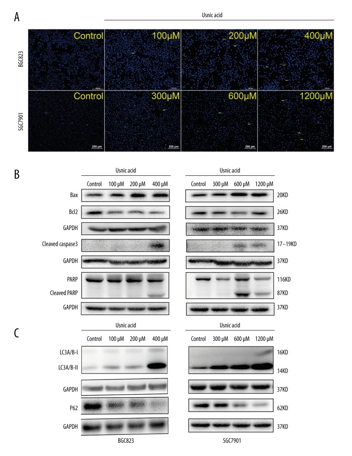
Figure 3.
Usnic acid (UA) induces cell apoptosis and autophagy in gastric cancer. (A) Hoechst (blue) staining: representative morphology and apoptotic cells (yellow arrows) of BGC823 and SGC7901 cells treated with UA for 24 h. Original magnifications: 40× (B) Expression of Bax, Bcl-2, cleaved caspase-3, and cleaved PARP in BGC823 and SGC7901 cells treated with UA at the indicated concentrations for 24 h as assessed by Western blot. (C) Western blot analysis of main cellular autophagic markers in the BGC823 and SGC7901 cells treated with UA at the indicated concentrations. Data show a concentration – response up-regulation of LC3 and down-regulation of p62 in cells treated with UA as compared to control. GAPDH was used as an internal control. All experiments were repeated independently at least 3 times.

