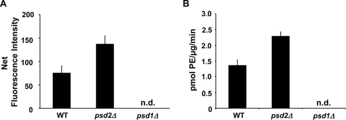
Figure 11.
Application of DSB-3–based fluorescence PSD assay to mitochondrial fractions from S. cerevisiae containing null mutations in PSD1 or PSD2. Mitochondrial fractions were obtained from the yeast strains of WT (BY4742) and psd mutant strains (psd1Δ::KanMX/BY4741 and psd2Δ::KanMX/BY4742) as described under “Experimental procedures.” Enzyme assays were performed with mitochondrial fractions (1 μg of protein/μl), 0.5 mm PS, and 3.1 mm Triton X-100 at 30 °C for 30 min. DSB-3 fluorescence detection with the PSD reaction products was conducted as described in Fig. 8. A, net fluorescence intensities of each PSD assay at the indicated time are shown after background correction. Background fluorescence is the value obtained from the PSD assay at zero time with heat-inactivated mitochondrial fractions. B, PSD activity of the mitochondrial fractions was calculated by applying the standard fluorescence data obtained as described in the legend to Fig. 8C. The data are from four independent experiments and are means ± S.E. (error bars). n.d., not detected.

