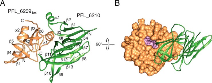
Figure 2.
Overall structure of PFL_6209tox–PFL_6210 complex. A, ribbon representation of PFL_6209tox (orange) in complex with PFL_6210 (green). Secondary structure elements and the location of the N and C termini of each protein are indicated. B, space-filling representation PFL_6209tox in complex with a ribbon representation of PFL_6210 rotated 90° with respect to A. Residues lining the cavity that extends into the core of PFL_6209tox are colored pink.

