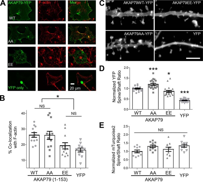
Figure 4.
Thr-87/Ser-92 phosphorylation disrupts F-actin binding in COS7 cells but affects synaptic targeting only mildly. A, AKAP79-YFP targeting domain wild-type and Thr-87/Ser-92 phospho-deficient (AA) and phospho-mimetic (EE) mutants or YFP alone expressed in COS7 cells (left), fixed, and Texas Red phalloidin-stained (middle), merged (right). B, percentage of co-localization of expressed AKAP79 with F-actin was determined using masks, as detailed under “Experimental procedures.” The phospho-mimetic (EE) AKAP79 (19.3 ± 2.0%) was significantly less co-localized to F-actin (p < 0.001, one-way ANOVA, Newman-Keuls post hoc analysis) than both WT (26.2 ± 1.5%) and phospho-deficient (AA) (25.9 ± 2.6%), but it was not significantly more co-localized to F-actin than empty YFP (16.5 ± 1.3%). n = 11–12 cells in each condition. C, AKAP79-YFP (top left), AKAP79(AA)-YFP (bottom left), AKAP79(EE)-YFP (bottom right), and YFP alone (top right) expressed in DIV 12 hippocampal neurons. D, quantification of spine to dendritic shaft ratio (normalized to control) from experiments from four individual neuron preparations as determined by 10 masks per neuron (n = 19–20 neurons per condition). AKAP79(AA)-YFP (1.20 ± 0.05) had significantly higher localization (p < 0.001, one-way ANOVA, Newman Keuls post hoc analysis) to dendritic spines compared with AKAP79-YFP (1.00 ± 0.03), whereas AKAP79(EE)-YFP (0.86 ± 0.05) spine localization was significantly reduced relative to control. All AKAP79 constructs expressed showed greater spine localization than YFP (0.43 ± 0.02) alone. E, spine to shaft ratio of an mTurquoise2 cell fill (images not shown) was unaffected by any of the AKAP constructs. *, p < 0.05; ***, p < 0.001; NS, not significant.

