
Figure 6.
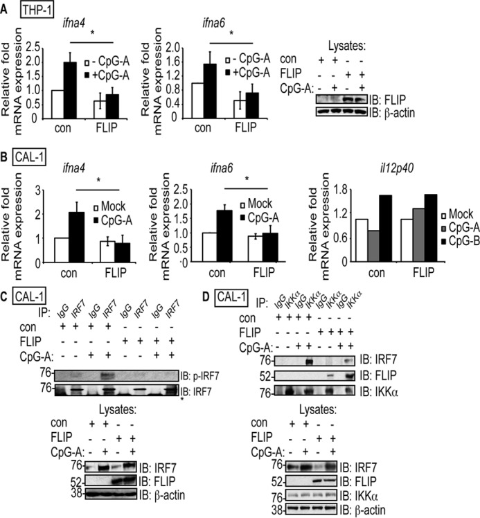
cFLIPL inhibits IRF7 activation and IKKα–IRF7 interactions in THP-1 and CAL-1 cell lines. A and B, THP-1 cells (A) and CAL-1 pDC cells (B) were transduced with a control lentivirus (con) or cFLIPL-expressing lentivirus (FLIP). THP-1 cells were differentiated into macrophages by treatment with PMA (10 ng/ml for 16 h). Transduced cells were incubated with medium lacking or containing 10 μm CpG-A or CpG-B for 5 h. Cells were lysed, and total RNA was extracted. A portion of each lysate was also used to detect cFLIPL protein expression. The levels of ifna4, ifna6, and il12p40 mRNA were quantified by using quantitative RT-PCR. C, transduced CAL-1 cells were incubated in medium lacking or containing 10 μm CpG-A for 5 h. Cells were then lysed, and a portion of lysate was immunoprecipitated with anti-IRF7 or nonspecific IgG antibodies. IB analysis of IP samples was performed to detect phospho-IRF7 or IRF7. A portion of each lysate prior to immunoprecipitation was also analyzed for expression of IRF7, cFLIPL, or β-actin. The asterisk denotes the heavy chain. D, transduced CAL-1 cells were incubated in medium lacking or containing 10 μm CpG-A for 5 h. Cells were then lysed, and a portion of lysate was immunoprecipitated with anti-IKKα or nonspecific IgG antibodies. IB analysis of IP samples was performed to detect endogenous IKKα, cFLIPL, or IRF7. A portion of each lysate prior to immunoprecipitation was also analyzed for protein expression.