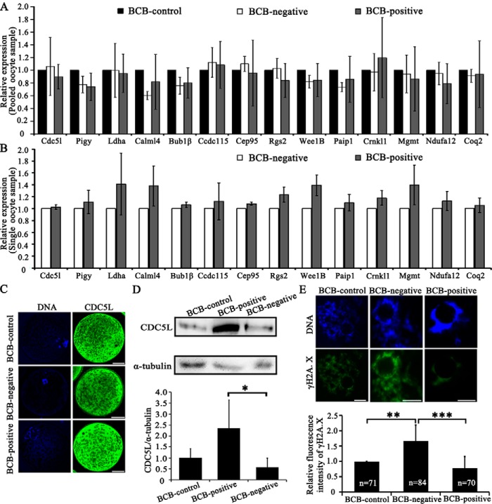
Figure 4.
Validation of single-oocyte RNA-seq results. A, RT-qPCR to validate 14 genes (cdc5l, pigy, ldha, calml4, bub1β, ccdc115, cep95, rgs2, wee1b, paip1, crnkl1, mgmt, ndufa12, and coq2) using pooled samples, which were selected from differential expressed genes identified by single-oocyte RNA-seq (details in Table S4). B, RT-qPCR using single-oocytes. The graphs presented the average relative expression levels of 9 single-oocytes (three replicates). C, immunostaining of the CDC5L protein, showing its localization in both the cytoplasm and nuclear area (except for nucleolus) of pig GV oocytes. Green, CDC5L; blue, DNA. Scale bar, 50 μm. D, Western blot of the CDC5L protein. Relative expression of CDC5L to α-tubulin in pig GV oocytes was quantified using ImageJ software. E, immunostaining of γH2A.X to indicate the occurrence of DSBs in GV oocytes. Relative fluorescence intensity of γH2A.X was quantified by ImageJ software. Green, γH2A.X; blue, DNA. Scale bar, 10 μm. *, p < 0.05; **, p < 0.01; and ***, p < 0.001.

