Figure 6.
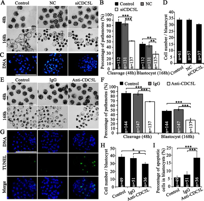
CDC5L vital to early embryo development. A, parthenotes (48 h) and blastocysts (168 h) after siCDC5L microinjection into oocytes at GV stages. Control, without microinjection; siCDC5L, cdc5l-specific siRNA group. B, total cleavage rates (≥2 cells at 48 h) and blastocyst rates (day 7) of parthenotes after siRNA microinjection. C, representative images of cell number of blastocysts. D, average cell number per blastocyst. E, parthenotes (48 h) and blastocysts (168 h) after antibody microinjection. Control, without injection. F, total cleavage rates (≥2 cells at 48 h) and blastocyst rates (day 7) of parthenotes after antibody microinjection. G, cell number and TUNEL signal of blastocysts at day 7 from control, oocytes injected with IgG and anti-CDC5L antibody. Green, TUNEL signal; blue, DNA. H, average cell number per blastocyst. I, percentages of apoptotic cells in blastocysts at day 7. *, p < 0.05 and ***, p < 0.001. Scale bar, 200 μm.

