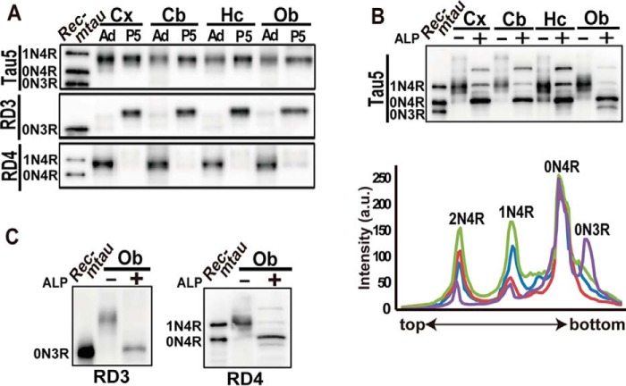
Figure 3.
Isoforms and the phosphorylation of tau in various brain regions of the adult mouse. A, immunoblots of tau in the cerebral cortex (Cx), cerebellum (Cb), hippocampus (Hc), and olfactory bulb (Ob) at P5 and at P90 (adult (Ad)) with Tau5, RD3, and RD4. The amounts of tau were adjusted with Tau5. Rec-mtau is recombinant mouse tau. B, an immunoblot of tau in four brain regions with Tau5 before (−) or after (+) dephosphorylation with ALP. Below are densitometric scans of dephosphorylated tau: blue, cerebral cortex; red, cerebellum; green, hippocampus; and purple, olfactory bulb. C, immunoblots of tau in the olfactory bulb (Ob) of adult mice with RD3 and RD4 after dephosphorylation with ALP (+). Tau before dephosphorylation is shown in lane −. The left lane is recombinant mouse tau (Rec-mtau).

