
Figure 7.
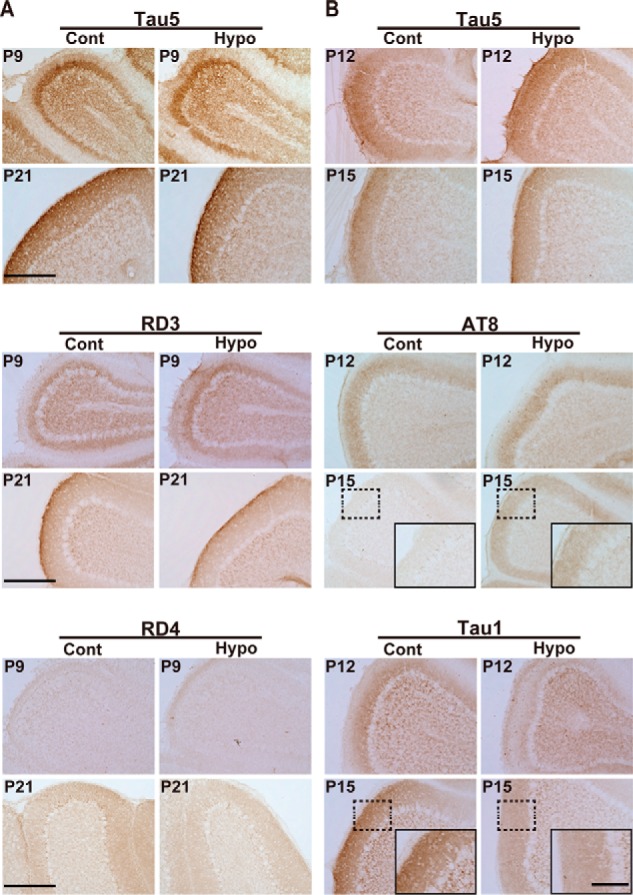
Immunohistochemical staining of mouse brains during postnatal development. A, cerebellum cortex of control (Cont) and hypothyroid (Hypo) mice at P9 and P21 stained with Tau5 (upper), RD3 (middle), and RD4 (lower). B, cerebellum cortex of control (Cont) and hypothyroid (Hypo) mouse at P12 and P15 stained with Tau5 (upper), AT8 (middle), and Tau1 (lower). Scale bar, 100 μm. Insets in AT8 and Tau1 at P15 are higher magnification of the region indicated by dotted box. Scale bar, 20 μm.