Fig. 2.
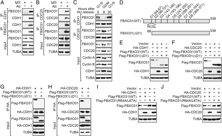
CDH1 and CDC20 interact with FBXO31 through its first D-box motif, which is required for proteasomal degradation. (A and B) Coimmunoprecipitation monitoring the interaction between endogenous FBXO31 and CDH1 (A) or CDC20 (B) in asynchronous HEK293T cells. IgG was used as a nonspecific control. (C) Coimmunoprecipitation monitoring the interaction between endogenous FBXO31 and CDH1 or CDC20 in HU-synchronized HEK293T cells 0 h (G1/S), 9 h (G2/M), or 20 h (G1) after HU release. (D) Schematic of WT FBXO31 showing the positions of the eight D-boxes, as well as the FBXO31(∆D1) and FBXO31(R64A,L67A) mutants. (E and F) Immunoblot monitoring Flag-FBXO31 levels (detected using an anti-Flag antibody) in HEK293T cells expressing Flag-FBXO31(WT) or Flag-FBXO31(ΔD1) and vector, HA-CDH1 (E), or HA-CDC20 (F). (G and H) Coimmunoprecipitation monitoring the interaction between ectopically expressed Flag-FBXO31(WT) or Flag-FBXO31(ΔD1) and HA-CDH1 (G) or HA-CDC20 (H) in HEK293T cells. (I and J) Immunoblot monitoring Flag-FBXO31 levels in HEK293T cells expressing Flag-FBXO31(WT) or Flag-FBXO31(R64A/L67A), and vector, HA-CDH1 (I), or HA-CDC20 (J).

