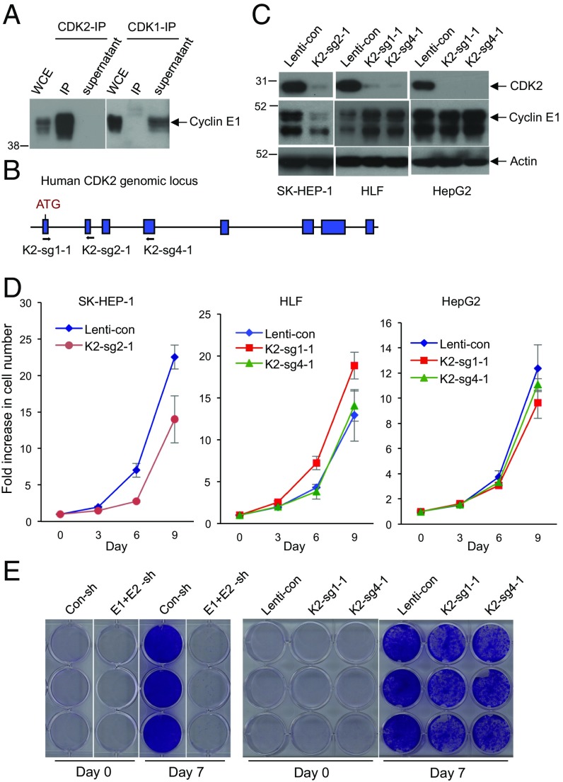
Fig. 3.
CDK2 is dispensable for HCC cell proliferation. (A) Immunoprecipitation (IP) using anti-CDK2 (Left) or anti-CDK1 (Right) antibodies, followed by Western blotting with an anti-cyclin E1 antibody. Note that immunoprecipitated CDK2 brings down large amounts of cyclin E1, and that there is essentially no cyclin E1 left in the supernatant after CDK2-IP. In contrast, no cyclin E1 was detected in CDK1-IP, and cyclin E1 remained in the supernatant after CDK1-IP. Whole cell extract (WCE) was also immunoblotted. (B) Design scheme of three independent guide RNAs against human CDK2. (C) Western blot analysis of CDK2 levels in three HCC cell lines after CRISPR-mediated knockout of CDK2 using three independent guide RNAs (K2-sg2-1, K2-sg1-1, and K2-sg4-1). Actin was used as a loading control. (D) Growth curves of SK-HEP-1, HLF, and HepG2 liver cancer cells following transduction with lentiviruses encoding K2-sg2-1, K2-sg1-1, or K2-sg4-1 guide RNAs (CDK2-knockout cells) compared with cells transduced with control viruses (Lenti-con). Error bars indicate SD, n = 3. (E) Comparison of HCC Huh-6 cell growth following knockdown of cyclins E1 and E2 (E1+E2-sh) versus after CRISPR-mediated knockout of CDK2 (K2-sg1-1 and K2-sg4-1). Con-sh and Lenti-con denote cells transduced with control shRNA or control-Lenti-viruses, respectively. Cells were fixed and stained with crystal violet 7 d after plating.

