Fig. 2.
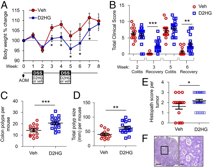
D2HG enhances tumorigenesis in the AOM-DSS mouse model of CAC. (A) Percent body weight change of mice i.p. injected with vehicle (red trace) or 25 mg/kg D2HG (blue trace) during DSS administration. (B) Clinical scores after DSS treatment and 1 wk recovery. (C and D) Number (C) and size distribution (D) of macroscopic polyps as determined using a dissecting microscope. (E) H&E-stained sections of Swiss-rolled colons were scored in a blind fashion for neoplasia. Four vehicle-treated mice did not have evidence of tumors when histopath scoring was performed. Results are presented as mean ± SEM of pooled data (A) or individual data points ± SEM (B–E) of 16 or 17 mice per group from three pooled, independent experiments. Two vehicle-treated mice and one D2HG-treated mouse did not survive until week 8 and are not included in sample size. *P < 0.05, **P < 0.01, ***P < 0.01 relative to vehicle, by one-way ANOVA followed by Bonferroni’s test (A and B) or unpaired, two-tailed Student’s t test (C–E). (F, Left) Photomicrograph of an intramucosal adenocarcinoma from a D2HG-treated mouse. Note the nuclear atypia, loss of basement membrane of glands, glandular crowding, and necrotic debris. (Scale bar: 200 μm.) (Right) Boxed area shown at higher magnification. (Scale bar: 100 μm.)

