Fig. 3.
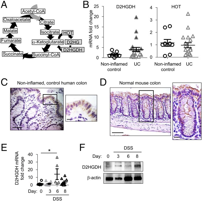
D2HGDH is predominantly expressed in the colonic epithelium and is increased during colitis. (A) D2HG formation in the tricarboxylic acid cycle. (B) Colonic D2HGDH and HOT mRNA expression measured by qPCR in human mucosal biopsies. Shaded triangles indicate a greater-than-threefold increase. (C and D) Immunohistochemistry staining of D2HGDH (brown stain) in human colonic mucosal biopsies (C) or mouse colon (D). (Scale bars: 50 μm.) Boxed areas are shown at higher magnification at right. (Scale bars for higher magnification images are 10 μm.) (E and F) D2HGDH mRNA expression measured by qPCR (E) and representative Western blot of D2HGDH protein expression (F) in isolated colonic epithelial cells from DSS-treated mice. Results in B and E are presented as individual data points ± SEM of nine normal patients or 21 UC patients (B) or five to nine mice per time point (E). *P < 0.05 by one-way ANOVA followed by Bonferroni’s test (E).

