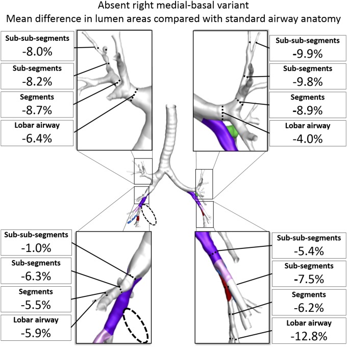
Fig. 3.
Mean differences in cross-sectional airway lumen areas by lobe and by anatomical level among participants with an absent right medial-basal airway branch variant (dashed circle) compared with standard anatomy in unaffected lobes. Airway lumen area comparisons are adjusted for lung volume. All mean differences were statistically significant (P < 0.01). See SI Appendix, Fig. S4 for standard airway anatomy lumen areas and mean differences with 95% CI.

