Fig. 4.
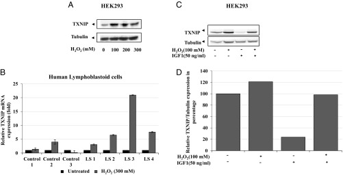
Effect of oxidative stress on TXNIP levels. (A) Serum-starved HEK293 cells were treated with H2O2 (100, 200, 300 mM) or left unstimulated. Lysates (100 μg) were analyzed by Western blotting for TXNIP and tubulin levels. (B) Effect of oxidative stress on TXNIP levels in LS-derived and control lymphoblastoids. Four individual LS and three control lymphoblastoid cell lines were treated with 300 mM of H2O2 or left unstimulated. Cells were harvested after 2 h and levels of TXNIP mRNA were measured by RT-QPCR. A value of 1 was given to TXNIP mRNA levels in untreated cells (solid bars). (C) Serum-starved HEK293 cells were treated with H2O2 (100 mM) or IGF1 (50 ng/mL) or both for 2 h. Lysates (100 μg) were analyzed by Western blotting. (D) Densitometric analysis of Western blot shown in Fig. 4C.

