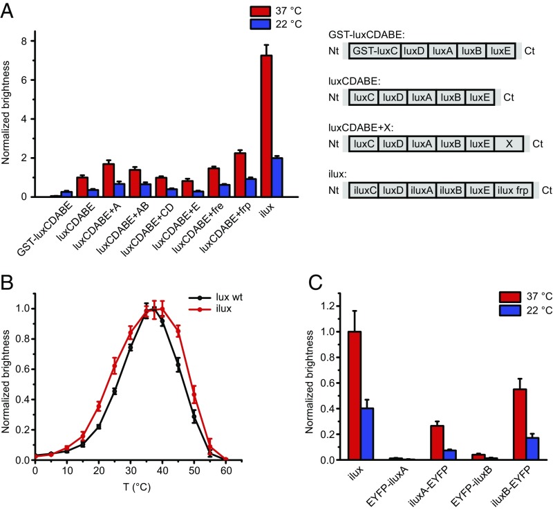
Fig. 1.
Generation and comparison of lux variants with increased brightness. (A) Brightness of different lux variants. The lux operons schematically shown on the Right were expressed from the vector pGEX(−) in DH5α cells on LB agar plates at 37 °C. Plates were imaged at 37 °C and 22 °C. Error bars represent SDs of six different clones. Nt, N terminus; Ct, C terminus. (B) Temperature curves of luxCDABE WT and ilux. The bioluminescence signal from DH5α cells expressing luxCDABE WT or ilux was measured in suspension at various temperatures. Error bars represent SDs of six independent measurements. (C) Brightness of ilux with EYFP-tagged versions of luxA and luxB. EYFP was introduced into ilux pGEX(−) at the N and C termini of luxA and luxB separated by a glycine-serine linker. Brightness was measured in DH5α cells on LB agar plates and normalized to the brightness of unlabeled ilux at 37 °C. Error bars represent SDs of four different clones.

