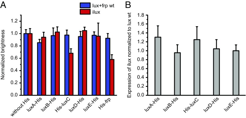
Fig. 2.
Brightness and expression of His-tagged proteins in luxCDABE+frp and ilux. (A) Brightness of luxCDABE+frp and ilux expressed from pGEX(−) in DH5α cells. A His tag with a glycine-serine linker was introduced into the indicated proteins in the lux operons. Brightness was measured on LB agar plates and normalized to the corresponding nontagged lux operon. Error bars represent SDs of 10 different clones. (B) Expression of His-tagged lux proteins. Whole-cell lysates of the same clones as in A were analyzed by Western blot. The His signal of each clone was normalized to the housekeeping protein DnaK. Subsequently, the ilux signal was normalized to the signal of the same protein in the luxCDABE+frp WT operon. Error bars represent SD.

