Fig. 1.
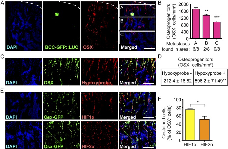
Breast cancer cells disseminate near hypoxic osteoprogenitor cells. (A) Immunohistochemistry showing GFP-expressing breast cancer cells (BCC-GFP::LUC) in green and OPCs, detected by anti-OSX immunostaining in red, in a wild-type mouse hind limb 5 d after i.c. injection. Dashed lines indicate the limit between the cartilage growth plate (above the dashed line) and the bone (below the dashed line). (B) Quantification of OSX+ cells and distribution of bone metastases in three areas (A, B, and C) below the growth plate cartilage; n = 8 independent bone metastases obtained from three mice 5 d after i.c. injections. (C) Immunohistochemistry showing that OSX expression (in green) colocalizes with hypoxia (Hypoxyprobe, in red) in hind limb sections of a wild-type mouse. (D) Quantification of OSX+ cells costained with Hypoxyprobe, showing that a majority of OPCs are hypoxic; n = 3 mice. (E) Immunohistochemistry showing that GFP expression driven by the Osx promoter (Osx-GFP, which marks OPCs, in green) colocalizes with HIF1α (in red; E, Upper Row) and HIF2α (in red; E, Lower Row) in the hind limbs of Osx-Cre::GFP transgenic mice. (F) Quantification of OSX+ cells expressing HIF1α or HIF2α; n = 3 mice with two sections per mouse. (Scale bars: 200 µm in A; 100 µm in C and E.) Values indicate the mean ± SEM, *P < 0.05, **P < 0.01, ****P < 0.0001, two-tailed Student’s t test.

