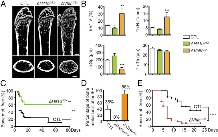
Fig. 2.
HIF signaling in osteoblast-lineage cells increases bone mass and bone metastasis. (A) Representative micro-CT images of sagittal (Upper) and transversal (Lower) views of distal femurs of control (CTL), ΔHif1αOSX, and ΔVhlhOSX mice. (B) Histomorphometric analyses of trabecular bone volume over tissue volume (BV/TV), trabecular number (Tb.N), trabecular separation (Tb.Sp), and trabecular thickness (Tb.Th) in control (n = 9), ΔHif1αOSX (n = 5), and ΔVhlhOSX (n = 3) femurs. (C) Kaplan–Meier analyses of bioluminescence-based detection of bone metastases after i.c. injections of BCC-GFP::LUC in control (n = 42) and ΔHif1αOSX (n = 27) mice. (D) Histological detection of bone metastases 30 d after i.f.p. transplantation of BCC-GFP::LUC in control (n = 12), ΔHif1αOSX (n = 6), and ΔVhlhOSX (n = 6) mice. (E) Kaplan–Meier analyses of bioluminescence-based detection of bone metastases after i.c. injections of BCC-GFP::LUC in control (n = 34) and ΔVhlhOSX (n = 23) mice. (Scale bars: 500 µm.) Values indicate the mean ± SEM, *P < 0.05, **P < 0.01, ***P < 0.001, ****P < 0.0001, two-tailed Student’s t test (B) or log-rank (Mantel–Cox) test (C and E).

