Figure 4.
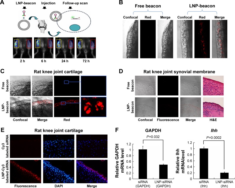
siRNA could be delivered into cartilage cells by LNPs and functioned in vivo.
Notes: Fluorescent signals could be detected by fluorescence molecular tomography in the LNP-beacon-treated mouse knee joint (red rectangles). In contrast, no fluorescent signal could be detected in the free-beacon-treated mouse knee joint (yellow rectangles) (A). The LNP-beacon-treated cartilage was further observed by confocal microscopy, and red signals were located in all of the cartilage tissue layers. In contrast, no signal was detected in the free-beacon-treated cartilage; magnification ×10 (B). Similar to mice cartilage data, red signals were present in the LNP-beacon-treated rat knee joint cartilage, whereas no signal was found in the free-beacon-treated rat knee joint cartilage; magnification ×10 for panels on the left and magnification ×20 for the insets (C). No positive signal could be detected by confocal microscope from either LNP-beacon-treated or free-beacon-treated rat knee joint synovial membrane; magnification ×10 (D). Fluorescent signals could be detected by fluorescence microscopy in the LNP-Cy3-labeled siRNA-treated rat knee cartilage. In contrast, no fluorescent signal was detected in the free Cy3-labeled siRNA-treated rat knee cartilage; magnification ×10 (E). After 24 hours of in vivo treatment with LNP-GAPDH siRNA or LNP-Ihh siRNA, cartilage GAPDH mRNA decreased by 51.8% and Ihh mRNA decreased by 79.7% (F).
Abbreviations: GAPDH, glyceraldehyde-3-phosphate dehydrogenase; Ihh, Indian Hedgehog; LNP, lipid nanoparticle; siRNA, small interference RNA.

