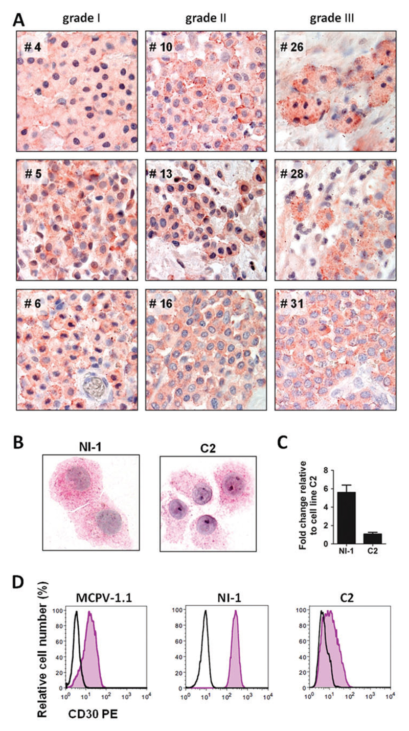
Figure 1.
Expression of CD30 in neoplastic canine mast cells. (A) Immunohistochemical detection of CD30 in primary neoplastic MCs in canine mastocytoma lesions. Tissue sections were prepared from formalin-fixed, paraffin-embedded tumour samples obtained from nine canine patients (left panels: grade I tumours: #4, #5, #6; middle panels: grade II: #10, #13, #16; right panels: grade III: #26, #28, #31). Sections were stained with monoclonal anti-CD30 antibody Ber-H2 as described in the text. Original magnification: ×100. (B) Immunocytochemical detection of CD30 in the canine mastocytoma cell lines NI-1 and C2 using anti-CD30 antibody Ber-H2. NI-1 and C2 cells were spun on cytospin-slides and expression of CD30 was analysed by immunocytochemistry. Images were taken using an Olympus microscope as described in the text. Original magnification: ×100. (C) CD30 mRNA expression in NI-1 and C2 cells. RNA isolation, cDNA synthesis and qPCR analysis were performed as described in the text. Expression levels of CD30 mRNA were calculated by the 2−ΔΔCT method and beta-actin was used as internal control. The figure shows the mean ± SD of three independent experiments. (D) CD30 surface expression on MCPV-1.1, NI-1 and C2 cells was analysed by flow cytometry using phycoerythrin (PE)-labelled monoclonal antibody BerH8 directed against CD30 (pink histograms). The isotype-matched control antibody is also shown (black open histograms). [Colour figure can be viewed at wileyonlinelibrary.com]

