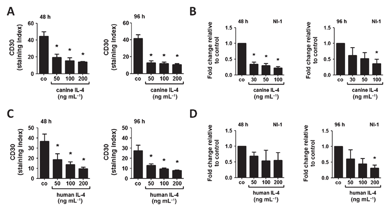
Figure 3.
Effects of canine and human IL-4 on CD30 expression in NI-1 cells. NI-1 cells were incubated with various concentrations of recombinant canine IL-4 (A, B) or recombinant human IL-4 (C, D) at 37 °C for 48 or 96 h. After incubation, expression of CD30 was determined by flow cytometry using anti-CD30 mAb BerH8; and expression of CD30 mRNA by qPCR analysis as described in the text. Flow cytometry results are expressed as staining index (= ratio of mean fluorescence intensity obtained with anti-CD30 mAb and isotype-matched control mAb); and results represent the mean ± SD of three independent experiments. Expression levels of CD30 mRNA were calculated by the 2−ΔΔCT method and beta-actin was used as internal control. The figures show the mean ± SD of three independent experiments. Asterisk (*): P <0.05.

