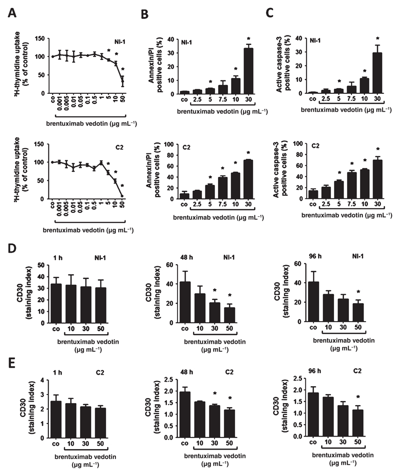
Figure 5.
Effects of brentuximab vedotin on proliferation, apoptosis and CD30 expression of NI-1 and C2 cells. (A) NI-1 and C2 cells were incubated with various concentrations of brentuximab vedotin at 37 °C for 96 h. Thereafter, 3H-thymidine uptake was measured. Results are expressed as percent of control (co) and represent the mean ± SD of three independent experiments. Asterisk (*): P < 0.05. (B, C) NI-1 and C2 cells were incubated in various concentrations of brentuximab vedotin at 37 °C for 96 h. Then, cells were examined by flow cytometry to determine the percentage of AnnexinV/PI-positive cells (B) and the percentage of active caspase-3 positive cells (C). Technical details are described in the text. Results represent the mean ± SD of three independent experiments. Asterisk (*): P < 0.05. (D, E) NI-1 and C2 cells were incubated in various concentrations of brentuximab vedotin at 37 °C for 1, 48 or 96 h. Thereafter, surface expression of CD30 was determined by flow cytometry. Results are expressed as staining index as described in the text. Results represent the mean ± SD of three independent experiments. Asterisk (*): P <0.05.

