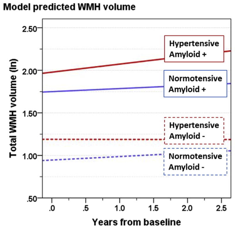
Fig. 1.

Using parameter estimates from Table 2, estimated trends of WMH volume as a function of years from baseline, are shown for prototype individuals for the following categories: normotensive/negative amyloid (blue, dashed), normotensive/positive amyloid (blue, solid), hypertensive/negative amyloid (red, dashed), and hypertensive/positive amyloid (red, solid). Amyloid positive threshold was CSF Aβ1–42 < 192 pg/mL; the amyloid level used in these calculations is the mean for each category. For each, the intercept represents a 74-year-old, APOE ε4 negative male with a high school education, no hyperlipidemia, and average intracranial volume. Abbreviation: WMH, white-matter hyperintensity. (For interpretation of the references to color in this figure legend, the reader is referred to the Web version of this article.)
