
Figure 1. Gastric mucosa. Also present: salivary, thyroid, kidney.
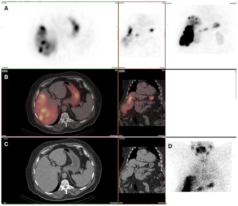
A 67-year-old male with hepatocellular carcinoma and no prior radioembolization was administered 99mTc-MAA via right groin access into the right hepatic artery and imaged 75 minutes after injection. Calculated lung shunt fraction was 14%. He underwent 90Y-radioembolization 15 days later without complication.
Axial, coronal, and MIP SPECT (Row A); Axial and coronal fused SPECT/CT (Row B); Axial and coronal CT (Row C); and anterior planar ( D) images demonstrate radiotracer uptake in the gastric mucosa. Note that the surrounding gastric serosal wall is spared, suggesting this is not related to an accessory artery arising from the hepatic vasculature. Expected radiotracer uptake is also seen in the right hepatic lobe tumor. Anterior planar images demonstrate uptake in the gastric mucosa as well as uptake in the salivary glands and thyroid gland. Note that gastric activity is uniform and diffuse, and spares the serosal gastric surface. Radiotracer uptake in the kidney (not shown) was also present. This uptake pattern, including gastric mucosa, salivary glands, and thyroid gland, is typical of free pertechnetate, and likely relates to in vivo breakdown of 99mTc-MAA.