
Figure 8. Gastric mucosa. Also present: salivary glands, thyroid, kidneys.
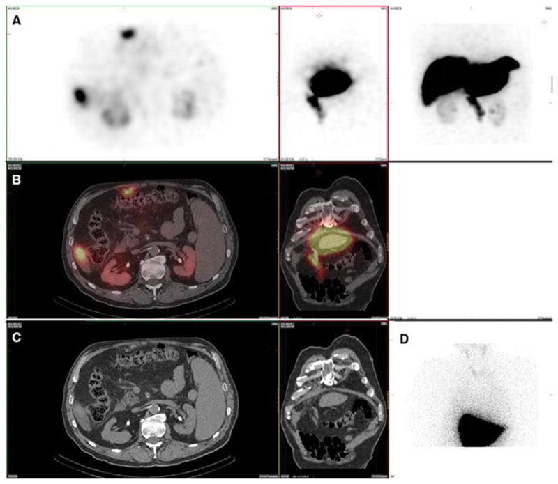
A 63-year-old male with metastatic neuroendocrine carcinoma and no prior radioembolization was injected with 99mTc-MAA via right groin access into the right hepatic artery and imaged 107 minutes after injection. Calculated lung shunt fraction was 8%. He underwent 90Y-radioembolization 21 days later without complication.
Axial, coronal, and MIP SPECT (Row A), axial and coronal fused SPECT/CT (Row B), axial and coronal CT (Row C), and anterior planar (D) images demonstrate radiotracer uptake throughout the gastric mucosa, but not in the gastric serosa, consistent with a pertechnetate distribution. Corresponding uptake in the salivary glands and thyroid gland is seen on the anterior planar image. Uptake was also seen in the kidney.